

Non-Contact Full-Field Microscopic Strain Measurement System

The VIC-3D Stereo Microscope system from Correlated Solutions is a highly accurate optical system for measuring surface shape, deformation, and strain on micro-scale specimens subject to an applied load. The high-magnification measurements are obtained by utilizing a stereo microscope with high-resolution machine-vision digital cameras, a custom optical beam splitter, a precision 3-axis motorized (or manual) stage, and patented software for complex distortion correction. This turnkey solution is ideal for measuring strain on composite fibers, solder joints, electrical components, bio materials, and much more.

Technology Background
Challenges Presented in Micro-Scale 3D DIC
Traditionally, three-dimensional DIC measurements have been difficult to obtain on specimens where high magnification is required. This is largely due to the lack of optics with sufficient depth-of-field to acquire two high-magnification images from different viewing angles. Stereo microscopes overcome the depth-of-field limitation, however, the complicated optics of using a single objective with a series of mirrors introduce distortions that are not accounted for in traditional calibration methods. These uncorrected images will result in severely biased shape and strain measurement data. In fact, it is not uncommon to observe bias levels of several thousand microstrain or more without performing a correction.
Solution: VIC-3D Stereo Microscope with Patented 3D Distortion Correction
To overcome this problem, Correlated Solutions has developed an easy-to-use calibration method that corrects for the complex, non-radial, optical distortions present in stereo microscopes. The VIC-3D Microscope calibration method computes and corrects for the non-parametric distortion fields of the stereo microscope, and has been shown to completely eliminate shape and strain bias from the measurements. Without this correction, measurement bias should be expected.

This exclusive procedure is essential when performing three-dimension DIC under a microscope. In this step, a sequence of images is taken of the provided speckled glass target. VIC-3D analyzes these image pairs to remove all optical distortions which are computed and corrected. With the motorized 3-axis precision stage, this distortion sequence becomes an automated process, saving time and further simplifying the calibration process.
Below are a few feature highlights of the VIC-3D Microscope system:
System Features

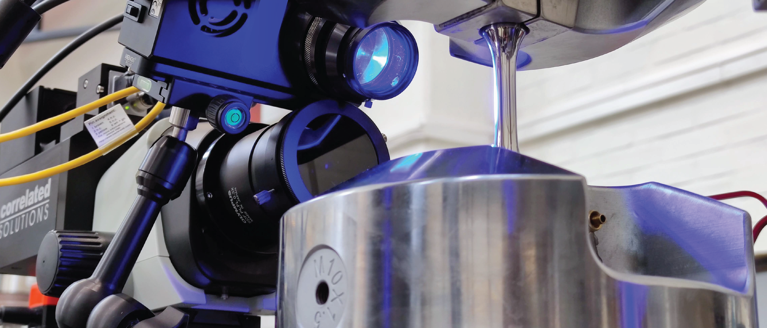
Typical VIC-3D Microscope System Lab Setup
Zoom Range (field of view): .8 mm to 7 mm
Full-field measurements of 3D coordinates, displacements, velocities, and complete strain tensors
Automatic image acquisition for distortion correction with a motorized stage
Perfect image overlap and parfocal focus/alignment for easy zooming between 1x-8x magnifications
Independent optical path adjustments for optimal image overlap
Powerful tools for visualizing data with iris
Contour displays can be overlaid onto images of the test specimen
Data extraction from 3D plots based on user-defined lines and circles
Convenient exporting of data
Data can be exported in Tecplot/plain ASCII, Matlab, and STL formats
Node data can be easily extracted for FEA validation
One-year of system maintenance included (unlimited technical support and software upgrades)
On-site support and consulting are also available
Two-year warranty for defects in materials and/or workmanship on all parts
Images courtesy of Chalmers University of Technology, Sweden (Department of Industrial and Materials Science)


